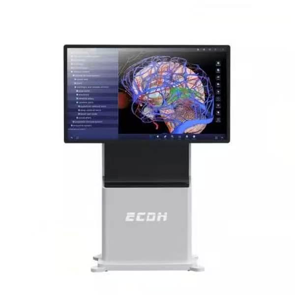
Anatomy departments are reaching a critical operational threshold. Specimen procurement costs rise annually, facility maintenance requires strict environmental controls, and expanding student cohorts strain existing laboratory resources — challenges that DIGIHUMAN addresses. Relying exclusively on physical wet labs creates an inherent bottleneck in medical curricula.
Universities require scalable methods to deliver precise anatomical instruction without inflating operational budgets. Virtual environments built on continuous tomographic data offer a functional solution. By replacing simplified 3D models with anatomically accurate digital human models, these platforms help address the persistent physical constraints of traditional medical training.
The Pedagogical Limits of the Wet Lab
Physical dissection provides undeniable tactile feedback, yet it contains structural limitations for group learning. A physical specimen degrades over time. Once a student cuts through a specific tissue layer, that structure cannot be restored for the next class. This single-use limitation restricts repeatable learning and minimizes the time each student spends analyzing complex spatial relationships.
Institutions also face heavy facility burdens. Maintaining cadavers requires specialized ventilation to manage harsh preservation chemicals like formaldehyde. Climate-controlled storage facilities incur ongoing energy demands. Biohazard disposal adds a recurring line item to department budgets. Universities need complementary systems that allow endless, non-destructive anatomical exploration across multiple student cohorts simultaneously.
Sub-Millimeter Precision Through Tomographic Data
Digital training systems only hold value if they match the accuracy of a real human body. High-end platforms are based primarily on real human tomographic data rather than simplified artistic models. Instead, they reconstruct 3D environments using actual continuous tomography from human subjects with no organic diseases or physical defects.
These databases are massive. The software relies on two distinct human data sets. The male data set contains 2,110 layers with sectional precision between 0.1mm and 1mm. The female data set pushes this accuracy further, featuring 3,640 layers with a sectional precision of 0.1mm to 0.5mm. By rendering these specific data points, the system reconstructs over 6,000 distinct anatomical structures.
The original pixels across these tomographic images exceed 1.2 billion. This extreme data density ensures that micro-vascular networks, fascia layers, and delicate nerve clusters remain clearly visible under heavy magnification. Faculty members can present microscopic structures without loss of visual clarity.
Standardizing Spatial Cognition Through Immersive Tech
Medical students frequently struggle to translate two-dimensional textbook diagrams into a three-dimensional understanding of the human body. Depth perception and spatial adjacent relationships are difficult to master through observation alone.
Hardware developers like DIGIHUMAN build solutions to target this specific learning gap. They utilize a helmet-type stereo interactive system to place the user directly inside the digital dataset. Students manipulate hand controllers to grab, rotate, and isolate specific organ systems from any angle. Sourcing human anatomy VR for institutions allows department heads to standardize this spatial training across an entire cohort. Real-time spatial tracking allows students to step inside organs and inspect internal cavities directly. If a user dissects the wrong vessel during a simulated exercise, the environment resets instantly. The classroom transforms into a controlled learning environment for surgical planning and repeated anatomical exploration.
High-Performance Rendering and Intuitive Software Control
Rendering billions of pixels in real-time requires immense computational power. In a live lecture setting, slow loading speeds ruin the instructional flow. If a professor has to wait two minutes for a digital skeletal system to load, student engagement drops.
To solve this, advanced virtual systems run on proprietary processing architectures. Powered by the self-developed Tai engine, these high-resolution platforms guarantee loading speeds of under 30 seconds. Instructors can transition instantly between the central nervous system and the cardiovascular system. The user interface includes jumping functions, multi-mode displays, and semantic association tools. Educators easily highlight target structures, hide surrounding tissue, and maintain total control over the lesson’s pace without network lag or system crashes. The system maintains high performance without frame drops during intensive interactive demonstrations.
Expanding Assessment and Cross-Disciplinary Study
True clinical preparation requires crossing the bridge between gross anatomy and modern diagnostic imaging. Medical professionals must know how a 3D organ appears on a 2D hospital scan.
Modern digital dissection platforms incorporate over 1,700 actual CT and MRI images. Students view the 3D anatomical model side-by-side with the corresponding radiological scan. This dual-view interface trains the eye to recognize pathological abnormalities across different diagnostic mediums. The software also features human peel and see-through tools, allowing learners to observe how superficial layers connect to deep tissue.
To support independent study, the system includes more than 130 micro-class videos featuring real dissection animations. Instructors can also pull from a massive repository of digital practice questions to track cohort progress, evaluate diagnostic accuracy, and verify knowledge retention before clinical rotations begin.
Digital human data can significantly improve the way anatomy programs operate. Universities reduce their reliance on expensive, single-use physical specimens while expanding student access to pristine anatomical models. By implementing high-resolution virtual systems, institutions build a scalable, risk-free environment that directly prepares students for modern clinical practice.
